
Illuminating Cancer.
Changing the Future of Research.
The TIME Lab brings oncologists, surgeons, and scientists to the same bench. With direct access to human tumors from the operating room, we study cancer not from a distance — but from the patient.


The battlefield inside every tumor
Cancer doesn’t exist in isolation. Every tumor builds itself a neighborhood — a Tumor Immune Microenvironment (TIME) comprising immune cells, connective tissue, blood vessels, and chemical signals that shapes whether the cancer grows or is destroyed. The immune system has the power to eliminate cancer cells, but tumors are extraordinarily good at manipulating their surroundings: silencing immune sentinels, excluding killer T cells, and recruiting bystander cells to hijack our defenses. The TIME Lab was built to map this hidden ecosystem, disrupt its defenses, and find the combinations of biology and medicine that let the immune system do what it was designed to do.
Our Approach
The operating room as a
catalyst for our research.
Patients as partners in the fight
Most cancer research begins with cell lines grown in a dish. Ours begins with patients — people facing some of the most difficult gastrointestinal cancers to treat — who choose to donate their tissues and tumors to help us understand and defeat these diseases. Because three of our five principal investigators are surgeon-scientists, the TIME Lab has built a research program grounded in this partnership. Every slice culture, every immune profile, every discovery we make begins with a patient who wanted to contribute to something larger than their own treatment. That generosity is what makes our work possible — and what drives our ability to innovate.
Learn more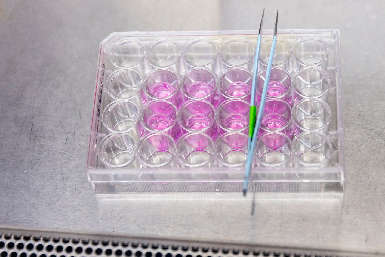
Our Platform
Tissue slice culture:
our platform for discovery
A living window into the tumor
Our lab pioneered a technique that changed how we study cancer: tumor slice culture. Instead of destroying a biopsy to extract cells, we keep thin sections of the tumor alive — intact, with all of its immune architecture preserved — for days in the lab. Inside these living slices, we can watch how immune cells move, test how tumors and immune cells respond to drugs, and interrogate the molecular conversations happening between cancer cells and the immune system in real time. It’s the closest thing to studying a patient’s tumor without the patient.
Read about Live Tissue Slice Culture in our Paper!Pancreatic Cancer
Pancreatic Cancer and the KRAS Puzzle
Overcoming barriers – translating to therapies
Pancreatic cancer has long been one of oncology’s hardest problems, and for decades, its most common driver — a mutation in a gene called KRAS — was considered simply undruggable. New molecules have finally broken that wall open. But our lab has identified a second problem: even when drugs can hit KRAS, T cells rarely reach the cancer cells to finish the job. They’re sequestered in the surrounding stroma, kept at arm’s length from the tumor by a microenvironment that was never supposed to let them in. We’re using our tumor slice culture platform to understand why — and to find the combination strategies that can let the immune system and these new targeted drugs work together.
Learn more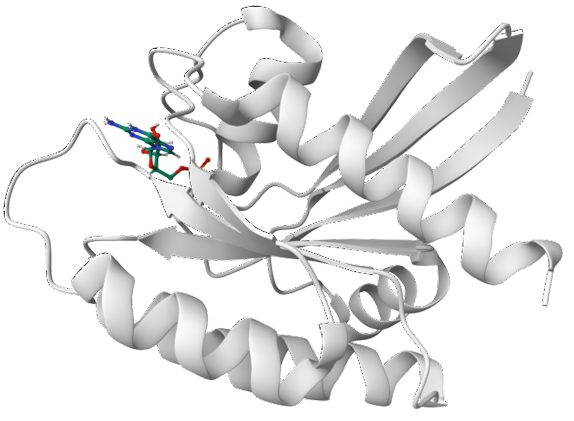
Pancreatic Cancer
Unlocking the tumor:
dual checkpoint blockade
Two locks, one key
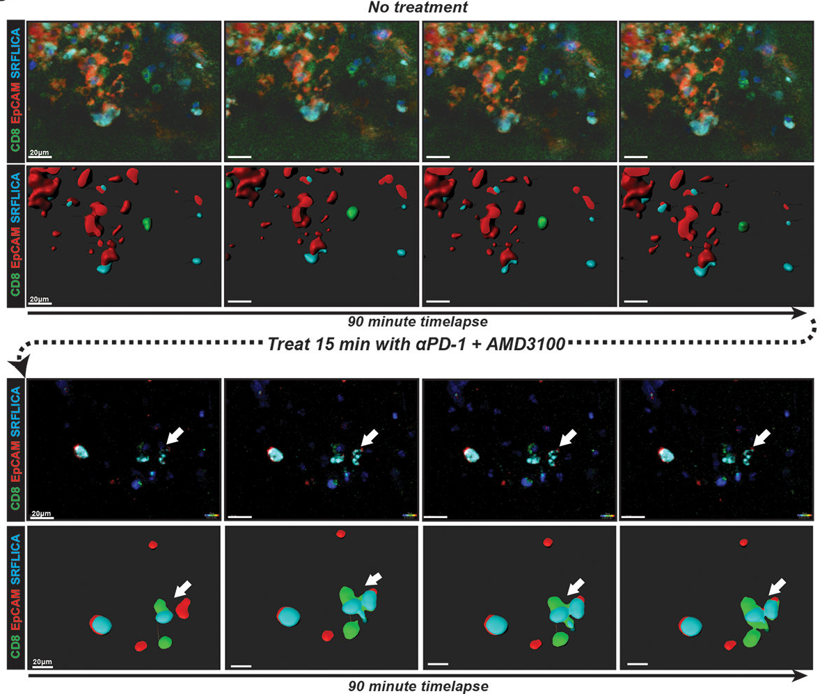
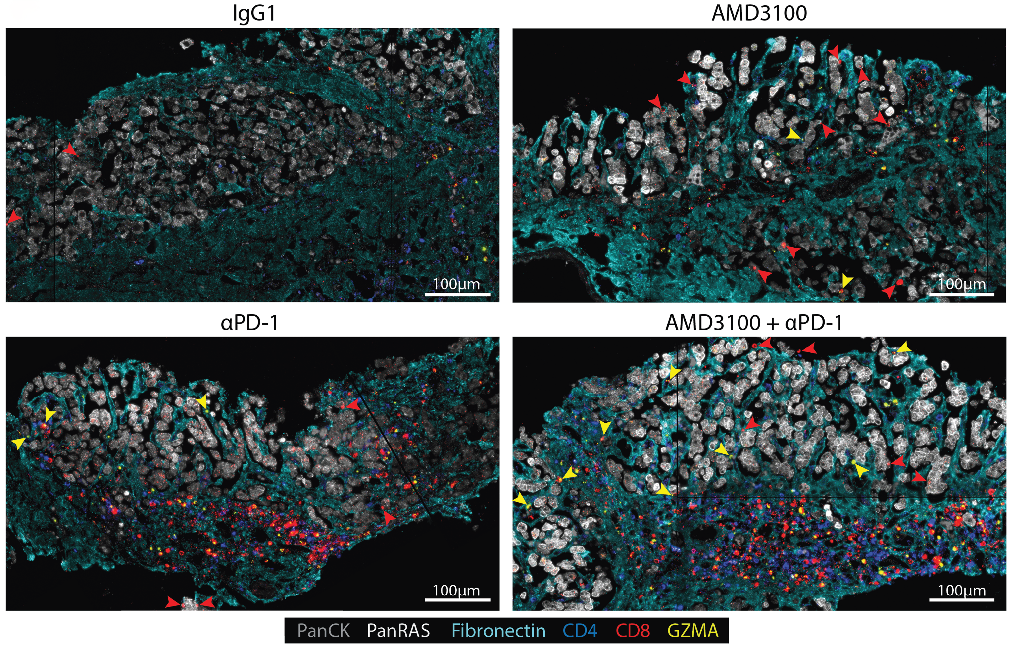
T cell exclusion from pancreatic tumors isn’t just a structural problem — it’s a chemical one. CXCR4, a receptor expressed on cancer cells and stromal cells alike, actively recruits immunosuppressive signals that reinforce the barrier keeping immune cells out. Our lab has shown that combining CXCR4 blockade with PD-1 checkpoint inhibition — attacking two different arms of the tumor’s immune evasion strategy simultaneously — can break this exclusion and allow T cells to reach and engage cancer cells in ways that neither drug achieves alone.
From bench to bedside
These findings have moved directly from bench to bedside. The TIME Lab is developing a clinical trial targeting this dual checkpoint axis, representing one of the first therapeutic strategies in pancreatic cancer specifically designed around our understanding of how the tumor immune microenvironment excludes T cells. For a disease with few treatment options, this approach opens a genuinely new door.
Learn moreColorectal Cancer & Liver Metastases
When cancer finds
a tolerant home
The liver's immunological blind spot
The liver evolved to be immunologically tolerant — protecting itself from overreacting to the constant flow of microbial signals from the gut. Colorectal cancer exploits exactly this quality. Once established in the liver, metastatic disease doesn’t just grow — it actively disables T cells, suppresses immune responses through molecules like IL-10, and enlists stromal cells to build barriers that keep the immune system at arm’s length. Our lab is working to reverse this suppression and restore the immune system’s ability to see and kill metastatic disease in the liver.
A problem that reaches beyond the liver
What makes liver metastases particularly dangerous is that their effects don’t stop at the liver’s edge. Evidence suggests that cancer established in the liver can disable T cell immunity to cancer elsewhere in the same patient — meaning that liver metastases may undermine the effectiveness of immunotherapy even for tumors in distant organs. Understanding how the liver’s tolerance machinery is hijacked by cancer, and how to interrupt it, is one of the most consequential questions in cancer immunology.
Learn more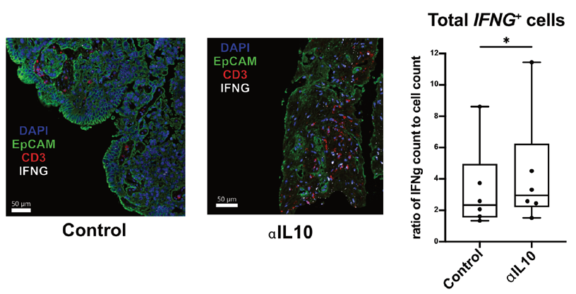
Fibrolamellar Carcinoma
Rare, young, and
no longer ignored
First answers for a forgotten disease
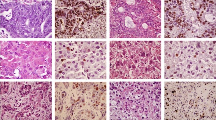
Fibrolamellar carcinoma strikes teenagers and young adults — people with no known risk factors and, until recently, no research community paying close attention. It is rare enough that most oncologists will never see a case, which has historically meant vanishingly little funding and even less mechanistic understanding. The TIME Lab is working to change that. Using our live tumor tissue platform, Drs. Damle, Pillarisetty, and Dr. Jason Carter are characterizing the immune environment of fibrolamellar tumors for the first time — asking whether the immunotherapy strategies reshaping treatment of other liver cancers can be brought to bear on a disease that has waited too long for answers. For patients with no approved therapies, these may be the first mechanistic data this disease has ever had.
Learn moreAppendiceal Cancer
A disease without
enough answers
The immune window we almost missed
Appendiceal cancer is rare, often misunderstood, and frequently diagnosed only after it has already spread throughout the abdomen — a condition called peritoneal carcinomatosis — because the original tumor causes so few symptoms. Patients are frequently treated with colon cancer protocols that may not fit their disease. Using a growing biorepository of more than 50 banked tumors and a library of patient-derived slice cultures, Dr. Sharib’s group is mapping the immune landscape of these peritoneal metastases from their earliest stages. Early findings suggest these tumors aren’t as immune-barren as once believed — and that the window for immune intervention may open earlier in the disease process than anyone expected. The very first foothold cancer gets in the peritoneum may carry an immune response that later disappears. Finding that window could change how we treat this disease entirely.
Learn more
Liver Immunology
The liver:
a unique immune frontier
Why the liver plays by different rules
No other organ in the body tolerates as much as the liver. It regenerates after injury. It resists T cell rejection after transplantation. It shelters malaria parasites from immune attack. And once cancer establishes itself there, it can disable T cell immunity not just locally — but throughout the body. The Crispe lab has spent decades asking why the liver’s immune rules are so different, and what happens when cancer learns to exploit them. Understanding this biology is essential to making immunotherapy work for the cancers that matter most to TIME Lab patients.
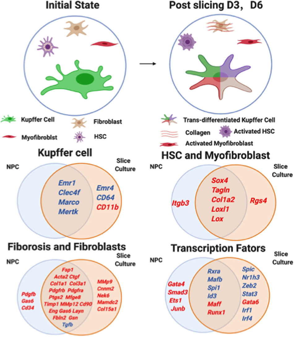
Kupffer cells and the origin of cancer-associated fibroblasts
Kupffer cells — the liver’s resident immune sentinels — are not a single population. Some originate in the yolk sac; others derive from bone marrow. A subset can trans-differentiate, taking on the characteristics of fibroblasts. The Crispe lab is investigating whether this plasticity is limited to yolk-sac-derived cells, and whether Kupffer cells drive the conversion of hepatic stellate cells into cancer-associated fibroblasts — the scaffolding that helps tumors exclude immune cells and resist therapy. The same mechanisms that drive liver fibrosis in chronic disease may be creating the conditions that allow liver metastases to thrive.
Learn moreTumor Immunology
Three cells. One
critical conversation.
The geometry of immune killing
Immunotherapy can eliminate cancer — but only in some patients, and oncologists still can’t reliably predict who will respond. The Damle lab has identified a key part of the answer: it isn’t just the number of immune cells inside a tumor that matters, but whether the right three cell types — a dendritic cell, a helper T cell, and a killer T cell — physically come together in the same place at the same time. These immune triads act as activation hubs: the dendritic cell presents tumor antigens, the helper T cell licenses the killer T cell, and together they orchestrate targeted cancer cell destruction. Using more than one million spatially resolved single-cell profiles across six assay platforms, we showed that these triads are a conserved feature of endogenous antitumor immunity — present even in notoriously immune-cold tumors like pancreatic cancer and fibrolamellar carcinoma — and that their density predicts patient survival.
Designing the assembly
Knowing that triads matter raises the next question: can we engineer them? In collaboration with the David Baker Lab at the UW Institute for Protein Design — home to the AI-driven protein design tools that have revolutionized structural biology — the TIME Lab is developing custom-designed binding proteins that physically bring these three cell types into contact. Rather than waiting for immune cells to find each other inside a hostile tumor microenvironment, these molecular scaffolds force the assembly of the triad, effectively engineering the spatial conditions that make immune killing possible.
Learn more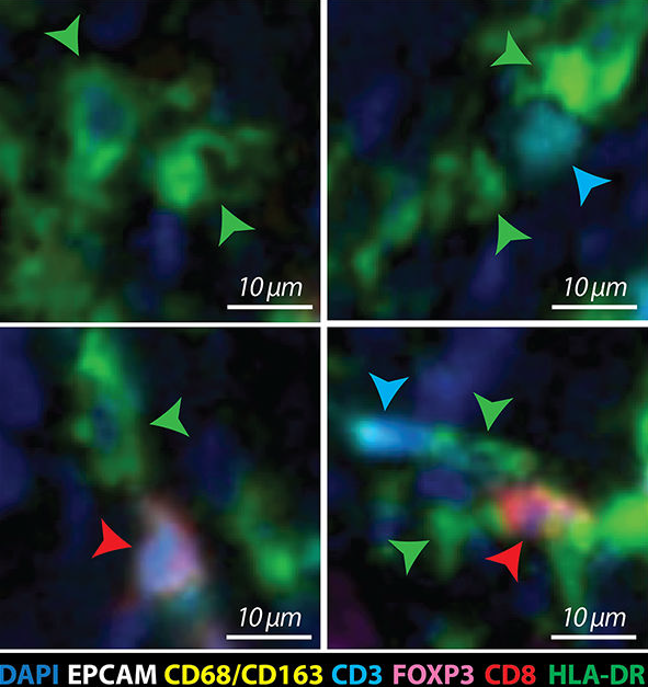
Team Science & Collaboration
Five labs. One shared purpose.
The TIME Lab is a highly collaborative, innovating research environment — three of its five principal investigators are surgeon-scientists, giving the group direct, privileged access to fresh human tumor tissue straight from the operating room. This proximity to patients doesn’t just shape what we study; it shapes how we think. Basic discovery, preclinical modeling, and clinical translation happen in the same building, between the same people, on overlapping timelines. The result is science that moves faster — and stays grounded to the questions that matter most.
A multi-PI consortium spanning surgical oncology, liver immunology, and translational biology — all under one roof at UW & Fred Hutch.
Operating room access means we study fresh patient tumors — not cell lines — bringing translational fidelity few labs can match.
A growing biorepository of annotated patient tumors — the foundation for collaborative science and personalized therapeutic discovery.
One question drives every experiment:
what would it take to let the immune system win?
The TIME Lab is committed to training the next generation of physician-scientists while driving research from the bench to the bedside. If you are interested in collaboration, training, or supporting our work, we’d love to hear from you.